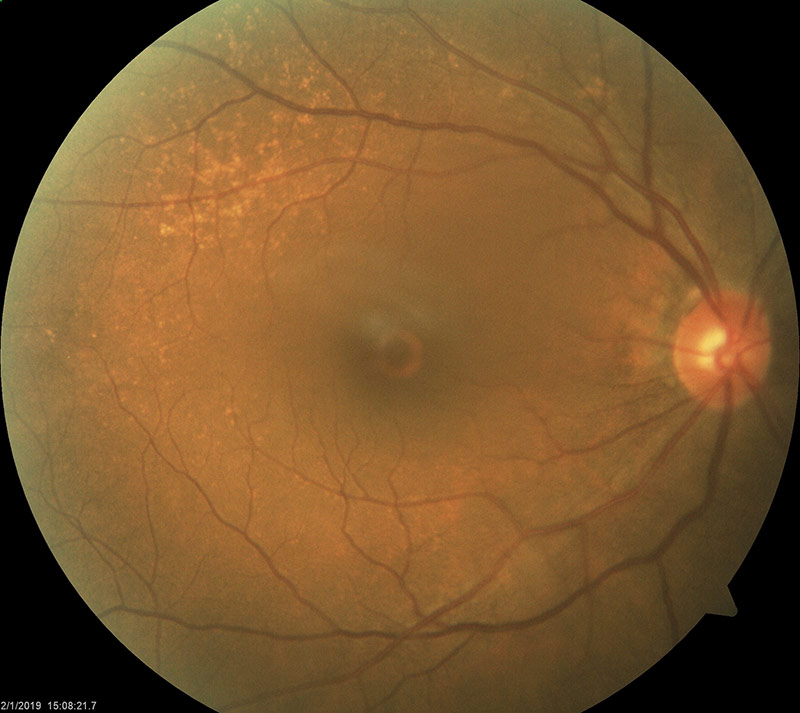
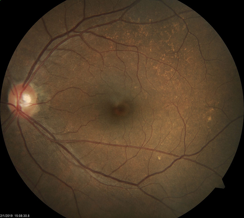
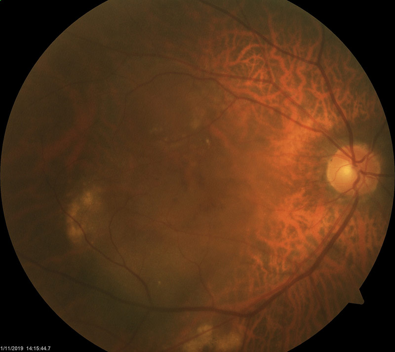
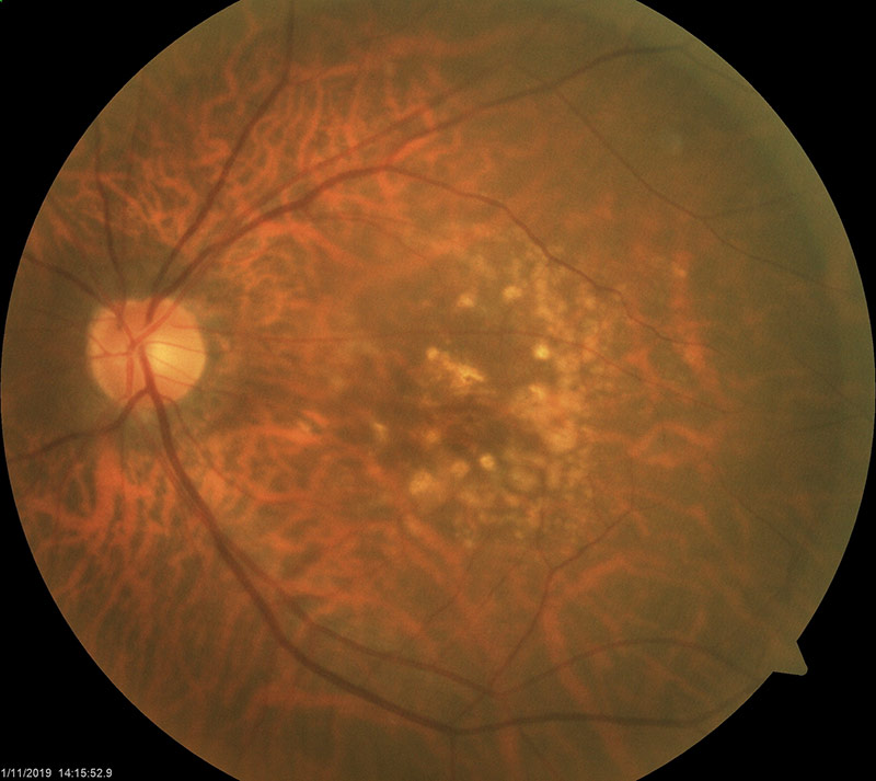
What is the macula?
The back of the eye is lined by the retina, which acts like a “film in a camera”, which is important in how we see.
The macula is a small area at the centre of the retina, which is essential for central and sharp vision. Abnormalities in the macula may result in the inability to see what is straight ahead though peripheral vision still remains good.
What is Age-Related Macular Degeneration (AMD)?
Age-related macular degeneration (AMD) is one of the leading causes of severe, irreversible vision loss in the world. It usually affects people aged 50 years and above as a natural result of aging. The cells in the macula become damaged and stop functioning effectively.
Visual loss from AMD can vary from mild to severe and has the tendency to affect both eyes.
Types of AMD
Dry AMD
Dry AMD occurs when the eye’s waste products are deposited in the region of the macula. This causes the retinal cells to degenerate. Although there is no effective treatment, the risks of vision loss are low unless the disease has reached a very advanced stage. This is the more common form of macular degeneration. It develops slowly over a number of years.


Wet AMD
Wet AMD is the result of abnormal blood vessels developing under the macula area. These are fragile and bleed easily, causing fluid or protein leakage and scarring. Unlike dry AMD, various forms of treatment are available but the loss of vision is often severe and permanent.


How is my sight affected by AMD?
Typically, only one eye is affected at first. In the early stages, it is possible that you may not notice the symptoms in one eye.
Symptoms of AMD
- An abnormality of the central part of your vision, in particular the appearance of a black patch or dark spot.
- Straight lines appearing wavy.
- You often misjudge distances and heights.
- Shades of colour appear “off”.
- You may need bright lighting conditions to see clearly.
- You have trouble with activities that require detailed vision, e.g. sewing.
- You may find letters of a word missing while reading.
- You may see the outline of a face but not the features.
How will my eye be examined for AMD?
When you come in for your outpatient appointment, you will have a sight test and then a full eye examination to determine the type of AMD you have.
Your ophthalmologist will instill drops into your eyes to make the pupils bigger, allowing them to see the retina and examine the back of your eye in detail. You will find that these eyedrops temporarily blur your vision. Because of this, your eyes may be sensitive to light and you may have difficulties reading. You should also therefore avoid driving on the day of your appointment.
You may also need one or more of the following further tests:
- An Amsler grid test, in which you look at a test page to check for blind spots.
- Colour photographs of the back of the eye taken with a special camera.
- A fluorescein or indocyanine green angiogram.
A small injection of fluorescent yellow or green dye is injected into a vein in your hand or forearm. As the dye circulates through the retinal blood vessels, abnormal areas in are lit up. This indicates that the vessels are diseased or new vessels have developed.
What treatment options are available?
If AMD is detected early, there are better chances for treatment. Depending on the form of the disease, vision can be improved or deterioration arrested.
Since there is no cure for AMD, early detection is important. People who have a family history of AMD should have an annual eye screening.
Treatment for Dry AMD
Supplements containing selenium, zinc and vitamins A, C and E may be prescribed. While they cannot cure AMD, nor restore vision, they may play a role in helping people with dry AMD at high risk from developing wet / advanced AMD.
Treatment for Wet AMD
Eye injections
Intravitreal Anti-Vascular Endothelial Growth Factors (anti-VEGF) injections have been observed to be successful in the treatment of Wet AMD. Such drugs are able to block the new vessel formation as well as associated swelling of the macular. These drugs typically need to be injected into the eye on a monthly basis. They have a 95% success rate in preventing loss of vision, and have a 30-40% success rate of improving vision. The effectiveness of these drugs will vary with the individual, depending on their use in the course of the disease. The risks of intravitreal injections include infection (approximately one in 3000 cases) and a low risk of stroke.
Laser therapy
A high energy laser beam is used to destroy the fragile, leaky blood vessels. Laser treatment may also damage some surrounding healthy retinal tissue. Hence, this method is only recommended if the leaky blood vessels have developed away from the central part of the macula as it can cause scarring that affects the vision.
Surgery
When there is bleeding directly beneath the macula, surgery can help treat the condition. Gas and a drug known as a tissue plasminogen activator are injected into the eye, moving the blood away from the macula and clearing the site.
